Page:EB1911 - Volume 27.djvu/391
are soft, painless swellings, sometimes of great size; though usually single, as many as a dozen may be present in the same individual . Lipomata are also found in the abdominal cavity, growing from the sub peritoneal layer of fat.

Fig. 5.—Lipoma of the palm.
What is known as a diffuse lipoma (fig . 6) consists of a generalized overgrowth of the subcutaneous fat of the neck, and this may be so extensive as to obliterate he outline of the jaw.

Fig. 6.—Diffuse lipoma of neck.
Fibroma (fig. 7).—Of tumours containing fibrous tissue, by far the most important are the fibroids of the uterus. A better name for these tumours would be Fibromyomata, as they always contain a varying proportion of muscle fibres. They originate in the wall of the uterus, but generally come to project either internally into the cavity of the uterus, or externally into the peritoneal cavity; and often their sole connexion with the uterine wall is a stalk or pedicle formed from the capsule of the tumour. Fibromyomata of the uterus are most common from 35 to 45 years of age; in girls under 20 they are almost unknown. They may attain a great size and are often multiple. They seem to be equally common in married and unmarried women. Not every fibroid is a source of danger or discomfort,

Fig. 7.—Uterus in sagittal section is showing interstitial and submucous fibroids.
for in the majority of cases they are discovered by chance or not until after death. On the other hand they may give rise to severe symptoms, and that in many different ways. First, they may cause haemorrhage prolonged over years so that the health is entirely ruined. Secondly, they may become inflamed and septic, and lead to severe blood-poisoning. Next, for some unknown reason, a fibroid tends to prevent conception, whilst, should pregnancy occur, labour is greatly impeded. Finally, it seems to be established that a fibroid may occasionally become converted into a sarcoma.
Examples of pure fibrous tissue tumours are the small multiple growths of the subcutaneous tissue, known as Painful subcutaneous nodule, and the irregular outgrowth from the gum known as Epulis.
A Myoma is composed of unstriped muscle fibres. It is a rare tumour sometimes found in the oesophagus, stomach and bladder.
Osteoma (fig. 8).—Bony tumours not infrequently arise from the bones of the head or face. They grow very slowly, and are so hard
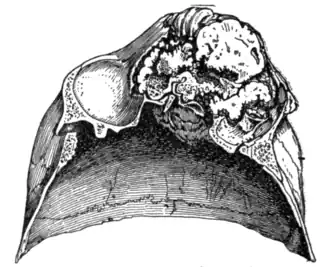
Fig. 8.—Osteoma of the left frontal sinus (seen from below).
that surgical removal may be very difficult. They also occur as irregular outgrowths from the bones of the limbs, and are then known as Exostoses (fig. 9). A common site for these is the inner and lower end of the femur, at the point of attachment of the adductor muscle, and such a tumour seems to originate from an ossification of the tendon of this muscle.

Fig. 9.—Exostosis of the femur produced by the ossification of the tendon of the adductor magnus.

Fig. 10.—Multiple chondromata of the fingers.
Chondroma (fig. 10).—Cartilaginous tumours are often found in children and young people growing from the bones of the limbs in the neighbourhood of the joints. They are frequently multiple, especially in the hands and feet. These tumours grow slowly and are quite painless. Should removal be necessary, it is usually an easy matter.
Odontoma.—Several varieties of this tumour have been described arising in connexion with the teeth and due to delayed or faulty development. They may cause great deformity of the jaw.
A Myxoma is composed of loose, gelatinous connective tissue similar to that found in the umbilical cord. Some nasal polypi seem to be of this nature, but true myxomatous tumours are rare. It is, however, not uncommon for a fibroma or a sarcoma to be converted by degeneration into myxomatous-like tissue.
Neuroma.—A pure neuroma is very uncommon, but a tumour known as a Pseudo-neuroma (fig. 11) is often found in the course of a nerve. This is formed by a localized overgrowth of the fibrous tissue of the nerve sheath.
Glioma.—This variety of tumour arises from the neuroglia, the