Page:EB1911 - Volume 25.djvu/188
mass is found which contains some remnants of the notochord. Elsewhere this structure is pressed out of existence and there is no further use for it when the cartilaginous vertebrae are once formed. One other series of structures must be mentioned though they do not
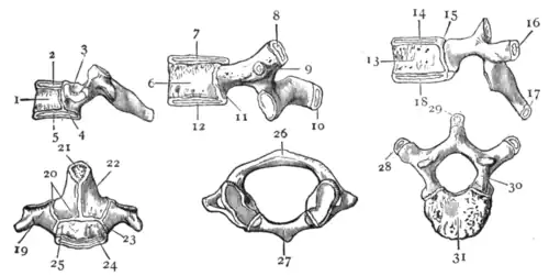
From Arthur Thomson, Cunningham's Text-Book of Anatomy.
Fig. 5.—Ossification of Vertebrae.
Cervical Vertebra.
1 Centre for body.
2 Superior epiphysial plate.
3 Anterior bar of transverse process developed by lateral extension from pedicle.
4 Neuro-central synchondrosis.
5 Inferior epiphysial plate.
Lumbar Vertebra.
6 Body.
7 Superior epiphysial plate.
8 Epiphysis for mammillary process.
9 Epiphysis for transverse process.
10 Epiphysis for spine.
11 Neuro-central synchondrosis.
12 Inferior epiphysial plate.
Dorsal Vertebra.
13 Centre for body.
14 Superior epiphysial plate, appears about puberty; unites at 25th year.
15 Neuro-central synchondrosis does not ossify till 5th or 6th year.
16 Appears at puberty; unites at 25th year.
17 Appears at puberty; unites at 25th year.
18 Appears about 6th week.
Axis.
19 Centre for transverse process and neural arch; appears about 8th week.
20 Synchondroses close about 3rd year.
21 Centre for summit of odontoid process; appears 3rd to 5th year, fuses 8th to 12th year.
22 Appears about 5th or 6th month; unites with opposite side 7th to 8th month.
23 Synchondrosis closes from 4th to 6th year.
24 Inferior epiphysial plate; appears appears about puberty, unites about 25th year.
25 Single or double centre for body; appears about 5th month.
Atlas.
26 Posterior arch and lateral masses developed from a single centre on either side, which appears about 7th week.
27 Anterior arch and portion of superior articular surface developed from single or double centre, appearing during 1st year.
28 Epiphysis for transverse process; appears about puberty, unites about 25th year.
29 Epiphysis appears about puberty; unites about 25th or 27th year.
30 Centre for neural arch on either side; appears about 6th or 7th week, the laminae unite from birth to 15th month.
31 Centre for body; appears about 6th week, unites with neural arch from 5th to 6th year.
play any great part in human development. In the intersegmental tissue ventral to each of the intervertebral disks a transverse rod of cells, known as a hypochordal bar, is formed which connects the heads of two opposite ribs. In man the greater number of these either disappear or form the middle fasciculus of the stellate ligament which joins the head of the rib to the intervertebral disk, but in the case of the atlas the rod chondrifies to form the anterior (ventral) arch which is therefore intersegmental, while the segmental body of the atlas, through which the notochord is passing, joins the axis to form the odontoid process. These hypochordal bars are interesting as the last remnant in man of the haemal arch of the vertebrae of fishes (see subsection on comparative anatomy). In the cervical region the ribs are very short and form the ventral boundary of the foramen for the vertebral artery. They are so short that little movement occurs between them and the rest of the vertebra, hence no joints are formed and the rib element becomes fused with the centrum and transverse process, leaving the vertebrarterial canal between. Sometimes in the seventh cervical vertebra the rib element is much longer and then of course more movement occurs, and instead of fusing with the rest of the vertebra it remains as a separate cervical rib with definite joints.
The sternum is developed according to G. Ruge by a fusion of the ventral ends of the ribs on each side thus forming two parallel longitudinal bars which chondrify and eventually fuse together in the mid line. The anterior seven or sometimes eight ribs reach the sternum, but the ventral ends of the ninth and sometimes the eighth probably remain as the xiphisternum, indeed a fibrous band is sometimes seen joining the caudal end of that structure to the ninth rib. The fusion of the two parallel bars begins at their cephalic ends and sometimes is interrupted toward the caudal end, thus leading to cleft or perforate sternum. At the cephalic end of each sternal bar, close to the place where the clavicles articulate, is an imperfectly separated patch of cartilage which usually fuses completely with the presternum, though sometimes it remains distinct and may later acquire a separate centre of ossification and so form a separate episternal bone on each side. If the sternum is to be regarded as the fused ventral ends of the thoracic ribs, the episternal elements are probably the remnants of the ventral ends of the seventh cervical ribs. The question of the morphological meaning of the sternum and surrounding parts cannot be settled entirely by a study of their development even when combined with what we know of their comparative anatomy or phylogeny. Professor A. M. Paterson (The Human Sternum, London, 1904) takes a different view from the foregoing and regards the sternum as derived from the shoulder girdle. To this point of view we shall return in the section on comparative anatomy.
The last stage in the development of the axial skeleton is the ossification of the cartilage; bony centres appear first in each half of the neural arches of the vertebrae and a little later (tenth week) double centres are deposited in the centra though these are so close together and fuse so rapidly that their double nature is often only indicated by their oval or dumb-bell-like appearance. The bone in the two halves of the neural arch spreads and fuses in the mid dorsal line, and later on joins the ossified centrum ventral to the facet for the rib. This point of junction remains as a narrow strip of cartilage for a long time and is known as the neuro-central suture or synchondrosis. The head of the rib therefore articulates with the developmental neural arch instead of the centrum. About the age of puberty secondary centres or epiphyses appear at the tips of the transverse and spinous processes and as thin plates just above and below the body (see fig. 5—2 and 3). These are fully united by the twenty-fifth year. In the lower two cervical vertebrae there is often a separate centre for the part corresponding to the rib, while the lumbar have an extra epiphysis for the mammillary process. The atlas has one centre for each side of the dorsal part of the arch and one (probably two fused) for the ventral part, which has already been referred to as a hypochordal bar. In the axis, in addition to the ordinary centres, there is one for each side of the odontoid process and one for the tip (see fig. 5—20, 21, 22). The sacral vertebrae have the usual centres, except that the anterior part of the lateral mass (costal element) has a separate centre and that there are two extra centres on each side of the whole sacrum where it articulates with the ilium (see fig. 6).
The ribs ossify by one primary centre appearing about the sixth week and by secondary ones for the tubercle and head. The sternum is ossified by centres which do not appear opposite the attachment of the ribs but alternately with them, so that although the original

From Arthur Thomson, Cunningham's Text-Book of Anatomy.
Fig. 6.—Ossification of Sacrum—a,a, Centres for bodies; b,b, Epiphysial plates on bodies; c,c, Centres for costal elements; d,d, Centres for neural arches; e,e, Lateral epiphyses.
cartilaginous structure is probably intersegmental the bony segments are segmental like those of the vertebral centra. As seven ribs articulate with the sternum six centres of ossification between them might be looked for, but there is so little room between the points of attachment of the sixth and seventh ribs that centres do not occur